“`
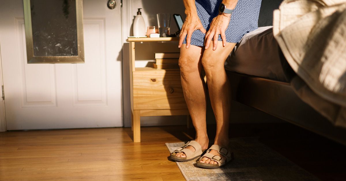
A knee that suddenly refuses to bend or straighten can feel as though someone slipped a doorstop into the joint. One moment you are walking, turning, or standing up; the next, your leg is stuck and every attempted movement starts an argument. This problem is commonly called a locked knee.
Knee locking is not a diagnosis by itself. It is a symptom that may come from a piece of tissue physically blocking the joint or from pain and swelling that make movement feel impossible. Some cases improve with rest and rehabilitation. Others, especially a knee that remains mechanically stuck after an injury, need prompt orthopedic assessment.
“`
What Does a Locked Knee Mean?
“`
A locked knee is an inability to move the knee through its normal range of motion. The person may be unable to fully straighten the leg, fully bend it, or both. The restriction can be constant or intermittent. Some people describe a sharp stop at one angle, while others report catching, clicking, or a momentary stuck feeling that releases after they shift position.
True knee locking
True locking occurs when something inside the joint creates a mechanical block. A displaced piece of torn meniscus is a classic example. A loose fragment of cartilage or bone can do the same thing. The knee may stop abruptly and refuse to move beyond a particular point, even when the person tries to relax.
Pseudo-locking
Pseudo-locking feels similar, but no fixed object is blocking the joint. Severe pain, inflammation, swelling, muscle spasm, or protective guarding makes the person unwilling or unable to move normally.
The distinction matters because true locking is more likely to require urgent imaging or a procedure. Pseudo-locking often improves when the underlying pain and inflammation are treated. Unfortunately, a knee does not display a convenient error message explaining which version you have.
“`
Common Causes of a Locked Knee
“`
1. Meniscus tears
The menisci are two C-shaped pads of tough cartilage between the thighbone and shinbone. They distribute pressure, absorb shock, and help stabilize the knee. A sudden pivot while the foot is planted can tear a meniscus, particularly during basketball, soccer, skiing, tennis, or an awkward squat.
In older adults, cartilage can weaken gradually. As a result, a minor twist, kneeling movement, or attempt to stand from a low chair may be enough to produce a degenerative tear.
A bucket-handle meniscus tear is especially associated with true knee locking. In this injury, a long strip of torn cartilage flips inward and may become lodged in the joint, preventing the leg from fully straightening. Other possible symptoms include joint-line pain, swelling, catching, popping, and the feeling that the knee may give way.
2. Loose bodies inside the joint
Small pieces of cartilage or bone can break free and float inside the knee. These fragments may move harmlessly for a while and then become trapped between the joint surfaces, causing sudden catching or locking.
Loose bodies can develop after an injury, from osteoarthritis, or because of an osteochondral injury involving both the cartilage and the bone beneath it. Symptoms may come and go as the fragment changes position, making the knee behave normally one hour and like a rusty folding chair the next.
3. Knee osteoarthritis
Osteoarthritis gradually damages the smooth cartilage that helps the knee glide. Rough joint surfaces, bone spurs, inflammation, and loose tissue can produce stiffness, grinding, catching, or a sticking sensation.
Arthritis-related locking is often intermittent. It may occur with morning stiffness, swelling after activity, reduced range of motion, and pain when climbing stairs or standing after sitting for a long time.
4. Osteochondritis dissecans
Osteochondritis dissecans occurs when a small area of bone beneath the joint surface loses adequate blood supply and begins to separate. The cartilage covering that area can loosen as well.
The condition is seen most often in children, teenagers, and young adults. If the bone-and-cartilage fragment becomes unstable, the knee may swell, give way, catch, or lock.
5. Kneecap dislocation or instability
The kneecap normally slides within a groove at the end of the thighbone. A blow, sudden change in direction, or structural alignment problem can cause it to slip partly or completely out of place.
Patellar instability may produce pain, swelling, buckling, catching, locking, or a visible change in the shape of the knee. A kneecap that remains dislocated requires prompt medical attention rather than a dramatic do-it-yourself shove.
6. Ligament injuries and major swelling
An anterior cruciate ligament injury or another significant knee sprain may not create a physical obstruction by itself. However, bleeding, swelling, pain, and muscle spasm can severely restrict movement and cause pseudo-locking.
A pop at the moment of injury, rapid swelling, instability, and difficulty putting weight on the leg can suggest a substantial internal injury. Meniscus and ligament injuries may also occur together.
7. Scar tissue and joint inflammation
Scar tissue after an operation or serious injury can restrict movement. This problem, sometimes called arthrofibrosis, may make the knee increasingly stiff and difficult to straighten.
An irritated synovial fold known as a plica can also cause pain, clicking, or catching. Inflammatory arthritis may create enough swelling and discomfort to make normal movement difficult, although it is less likely to produce a fixed mechanical block.
8. Joint infection and other uncommon causes
A joint infection can cause intense pain, warmth, swelling, and severe loss of motion. Tumors, cysts, crystal arthritis, and other unusual conditions can also interfere with knee movement.
These causes are less common than meniscus tears or arthritis, but they become important when symptoms occur without a typical injury or are accompanied by fever, redness, unexplained weight loss, persistent nighttime pain, or feeling seriously unwell.
“`
Locked Knee Symptoms to Watch For
“`
The defining symptom is reduced movement, but the surrounding details can help identify the likely cause. Common locked knee symptoms include:
- Inability to fully straighten or bend the knee
- A hard stop or blocked feeling at a particular angle
- Catching, clicking, popping, or grinding
- Pain along the inner or outer joint line
- Swelling, stiffness, or warmth
- Buckling or a sensation that the knee may give way
- Difficulty walking, climbing stairs, squatting, or standing from a chair
- Pain that becomes worse during twisting or pivoting
The timing of swelling may provide useful clues. Immediate, substantial swelling after trauma can signal bleeding inside the joint from a serious ligament, bone, or cartilage injury. Swelling that gradually develops over the next day or two may occur with a meniscus tear or irritation after activity.
These patterns are clues rather than reliable home diagnostic tests. Knee injuries are annoyingly talented at producing similar symptoms from very different problems.
“`
When Is a Locked Knee an Emergency?
“`
Seek urgent medical assessment when the knee remains stuck after an injury, particularly when you cannot straighten it or put weight on the affected leg. A truly locked knee may contain displaced tissue that needs timely treatment.
Prompt care is also important if you experience:
- Severe or rapidly worsening pain
- Rapid, extensive swelling
- An obvious deformity
- A kneecap that remains out of position
- Numbness or weakness below the knee
- A cold, pale, or bluish foot
- A loud pop followed by major instability
- Inability to take several steps safely
A hot, red, swollen knee accompanied by fever or chills can indicate a joint infection and requires same-day emergency evaluation. Do not force a deformed or firmly locked knee back into position. Forceful movement could worsen cartilage, ligament, nerve, or blood-vessel damage.
“`
How Doctors Diagnose Knee Locking
“`
Reviewing the medical history
A healthcare professional will ask how the problem started, whether the knee twisted or received a direct blow, and how quickly swelling appeared. The clinician may also ask whether the knee catches at the same angle every time or whether movement is mainly limited by pain.
Age, sports participation, occupation, earlier injuries, arthritis, previous operations, medication use, and systemic symptoms can help narrow the possibilities.
Performing a physical examination
The examination usually includes checking for swelling, bruising, warmth, tenderness, and deformity. The clinician will gently test how far the knee can bend and straighten and may examine the kneecap’s position and movement.
When pain permits, ligament stability tests may be performed. Maneuvers such as the McMurray or Thessaly test can help identify signs associated with a meniscus injury. However, these examinations are only pieces of the diagnostic puzzle, not magical buttons that immediately reveal what is wrong.
X-rays
X-rays can identify fractures, arthritis, alignment abnormalities, bone spurs, and some loose bodies. They do not show a meniscus tear clearly because the meniscus is made of soft tissue rather than bone.
Magnetic resonance imaging
Magnetic resonance imaging, or MRI, provides detailed images of the menisci, ligaments, cartilage, tendons, and bone marrow. It may be recommended when a displaced meniscus tear, ligament injury, cartilage defect, or another internal problem is suspected.
MRI can be particularly useful after a traumatic injury when a person cannot fully extend the knee. However, imaging findings must be matched with symptoms because some meniscus tears, especially degenerative tears in older adults, may exist without causing the current problem.
Additional tests
Ultrasound may help evaluate selected tendons, cysts, or fluid collections. Computed tomography may be useful when a complicated fracture or certain bone abnormalities need closer examination.
If infection or crystal arthritis is suspected, a clinician may remove fluid from the knee with a needle and send it for laboratory analysis. The appropriate test depends on the history and examination. Ordering every scan available is not a medical strategy; it is merely an expensive scavenger hunt.
“`
Locked Knee Treatment
“`
Locked knee treatment depends on whether the joint is truly blocked, which structure is injured, the severity of the symptoms, the person’s age and activity level, the amount of existing arthritis, and how long the problem has been present.
Safe first steps
Stop sports and avoid twisting, pivoting, kneeling, or deep squatting until the knee has been evaluated. Do not repeatedly test whether the joint has changed its mind.
Initial care may include:
- Resting and protecting the leg
- Applying a wrapped cold pack for about 15 to 20 minutes at a time
- Using light compression if it feels comfortable
- Elevating the knee above heart level when practical
- Using crutches if walking is painful or unstable
Never apply ice directly to the skin, and do not wrap the knee so tightly that the foot becomes numb, cold, or discolored.
Over-the-counter pain medicine may be appropriate for some people. However, allergies, stomach ulcers, kidney problems, blood thinners, pregnancy, age, and other medical conditions can change what is safe. Follow the product label and consult a healthcare professional when uncertain.
Nonsurgical treatment
Many knee problems respond to a structured conservative plan. Treatment may include temporary activity modification, a brace, medication when appropriate, and physical therapy.
Rehabilitation can help restore range of motion, strengthen the quadriceps and hip muscles, improve balance, and correct movement patterns that place excessive pressure on the knee. Progress should be gradual. Aggressively stretching a joint that is physically blocked is not recommended.
Small, stable meniscus tears may improve without surgery, especially when they do not cause persistent true locking. Degenerative tears associated with osteoarthritis are commonly treated first with exercise-based therapy and arthritis management rather than immediate arthroscopy.
Injections may occasionally reduce inflammation related to arthritis. They cannot repair a torn piece of cartilage or remove a loose body that is physically blocking the joint.
Arthroscopic knee surgery
Persistent mechanical locking from a displaced meniscus fragment or loose body may require knee arthroscopy. During this minimally invasive procedure, a surgeon places a small camera and specialized instruments into the joint through small incisions.
Depending on the cause, the surgeon may:
- Repair a torn meniscus
- Trim unstable, damaged meniscus tissue
- Remove a loose cartilage or bone fragment
- Treat a damaged cartilage surface
- Release restrictive scar tissue
Preserving healthy meniscus tissue is valuable because the meniscus protects the joint and distributes pressure. Whether a tear can be repaired depends on its location, size, pattern, blood supply, tissue quality, and associated injuries.
A meniscus repair generally requires a longer and more carefully protected recovery than trimming damaged tissue. However, successful repair may offer better long-term joint protection in suitable patients.
Treatment for knee arthritis
Arthritis treatment may involve low-impact exercise, strength training, weight management when relevant, braces, pain-relieving medication, and injections. Activities such as cycling, swimming, and controlled walking can maintain movement without the repeated impact of running.
Advanced arthritis that causes major pain, frequent locking, and substantial disability may eventually require partial or total knee replacement. This decision is usually based on symptoms and loss of function rather than an X-ray alone.
Treatment for kneecap instability
A first kneecap dislocation may be treated with temporary support, activity restriction, and rehabilitation after the kneecap has been safely returned to position. Physical therapy typically focuses on leg strength, hip control, flexibility, and movement mechanics.
Repeated dislocations or significant cartilage damage may require surgery to stabilize or realign the kneecap.
“`
Recovery and Prevention
“`
Recovery can range from several days for a mild pain-related restriction to several months after meniscus repair or another operation. Healing time depends on the exact injury, treatment, age, health, and rehabilitation progress.
A safe return to sports should be based on restored movement, strength, balance, and confidence rather than simply reaching a date circled on the calendar.
Not every locked knee can be prevented, but risk may be reduced by:
- Strengthening the quadriceps, hamstrings, hips, and core
- Increasing exercise intensity gradually
- Warming up before demanding activity
- Practicing safe landing and cutting techniques
- Wearing footwear suited to the activity and surface
- Avoiding sudden pivots on a firmly planted foot when possible
- Allowing adequate recovery after previous knee injuries
For people with osteoarthritis, consistent low-impact movement can help maintain mobility and muscle support. Doing too little may increase stiffness, while doing too much too quickly can trigger a painful flare. The useful middle ground is rarely glamorous, but knees generally prefer consistency over heroics.
“`
Experience-Based Perspective: What the Locked-Knee Journey Often Feels Like
“`
The following scenarios are composite examples created to explain common experiences. They do not describe specific patients and cannot replace a professional examination.
The athlete who tries to walk it off
A recreational soccer player plants one foot, turns to chase the ball, and feels a pop. The pain is noticeable but not dramatic, so the player finishes the game. By the next morning, the knee is swollen and will not fully straighten.
The surprising lesson is that being able to walk immediately after an injury does not rule out a meniscus tear. Adrenaline, momentum, and stubborn optimism can keep someone moving even when the joint has been damaged.
The most useful response is not to perform increasingly creative hallway lunges in hopes of unlocking the knee. It is to protect the joint, avoid pivoting, and arrange an assessment. If imaging confirms a displaced bucket-handle tear, the treatment discussion may move quickly toward arthroscopy because the fragment is physically blocking extension.
After treatment, rehabilitation can feel frustratingly slow at first. However, milestones such as reducing swelling, activating the quadriceps, restoring extension, and developing a smooth walking pattern matter more than dramatic daily gains.
The adult whose knee sticks after sitting
Another common experience is less cinematic. An adult with gradually increasing knee pain notices that the joint feels stuck after a long car ride or an evening on the couch. It loosens after several careful steps but catches again on stairs. There was no memorable injury, and X-rays show osteoarthritis.
In this situation, the sticking sensation may come from stiffness, inflammation, rough cartilage, or loose fragments rather than a fresh sports injury.
The turning point is often learning that rest alone is not a complete treatment plan. Too much inactivity can increase stiffness and weaken the muscles supporting the knee. A measured program of low-impact movement and strengthening may make daily tasks easier, even though it cannot return worn cartilage to factory condition.
Tracking which activities trigger swelling, using pacing rather than boom-and-bust exercise, and working with a physical therapist can be more productive than buying a drawer full of braces with heroic packaging.
The teenager with a shifting kneecap
A teenager may describe the kneecap sliding sideways during a quick change in direction, followed by pain, swelling, and fear that the knee will buckle again. Sometimes the patella returns to position on its own. In other cases, it remains visibly displaced and requires urgent medical reduction.
Even after the pain settles, apprehension can linger. The person may avoid bending the knee or stop trusting the leg during sports. Recovery therefore involves more than reducing swelling.
Strengthening the quadriceps, hips, and trunk, improving landing control, and gradually rebuilding confidence are central parts of rehabilitation. Recurrent instability deserves specialist evaluation because repeated episodes can damage cartilage.
The person whose symptoms disappear before the appointment
Some people finally schedule an appointment, only for the knee to unlock and behave perfectly in the examination room. This can feel embarrassing, but intermittent symptoms are still medically relevant. A loose body or unstable meniscus fragment can shift position, causing locking one day and nearly normal movement the next.
Writing down when the locking occurred, what movement triggered it, where the pain was located, how long the episode lasted, and whether swelling followed can help the clinician. A short video of the movement problem may also be useful when safely captured without recreating a painful maneuver.
The larger lesson across these experiences is simple: locked knee describes an experience, not one specific injury. The right solution comes from identifying why the joint is stuck rather than treating every case as though the same invisible wrench has fallen into the machinery.
“`
Conclusion
“`
A locked knee can result from a displaced meniscus tear, loose cartilage or bone, osteoarthritis, osteochondritis dissecans, kneecap instability, scar tissue, or severe pain and swelling after an injury.
A knee that remains mechanically blocked, becomes deformed, cannot support weight, or is hot and swollen with fever needs prompt medical attention. Until the cause is known, protect the joint and resist the temptation to force it straight.
Diagnosis usually combines the story of how the symptoms began, a careful physical examination, and selected imaging. Treatment may range from temporary protection and physical therapy to arthroscopic repair or removal of a mechanical obstruction.
The best outcome starts with recognizing that a stuck knee is a message from the jointand unlike most group chats, this is one message worth opening immediately.
“`