Porphyria sounds like the name of a mysterious kingdom in a fantasy novel. In reality, it is a group of rare metabolic disorders that can affect the nervous system, skin, liver, and other organs. Its symptoms may resemble everything from food poisoning and anxiety to nerve disease and severe sunburn, which explains why obtaining the correct diagnosis can feel like solving a medical mystery with several missing pages.
The good news is that porphyria can be identified with the right biochemical tests, and modern treatments can stop attacks, reduce future episodes, protect the skin, and help many people maintain active lives. The crucial first step is determining exactly which type of porphyria is present because the symptoms, triggers, tests, and treatments are not identical.
What Is Porphyria?
Porphyria is an umbrella term for disorders involving the production of heme, an iron-containing molecule best known as a component of hemoglobin. Heme also supports liver enzymes, energy production, medication metabolism, and many other cellular processes.
The body builds heme through a multistep biochemical pathway. Think of it as an assembly line with several specialized workers. If one enzyme does not perform its assigned step correctly, porphyrins or their chemical precursors can accumulate. Those substances may then injure nerves, irritate the skin when exposed to light, or affect the liver.
Most porphyrias are caused by inherited gene variants, although having a variant does not guarantee that symptoms will develop. Porphyria cutanea tarda, the most common cutaneous form, is often acquired or triggered by a combination of genetic susceptibility, excess iron, liver disease, alcohol use, smoking, estrogen exposure, or certain infections.
Acute porphyrias
Acute porphyrias primarily affect the nervous system and internal organs. Symptoms usually occur in attacks lasting days or, in severe cases, weeks. The four acute hepatic porphyrias are:
- Acute intermittent porphyria
- Variegate porphyria
- Hereditary coproporphyria
- ALA dehydratase deficiency porphyria
Variegate porphyria and hereditary coproporphyria may cause both acute neurologic attacks and blistering skin symptoms. Acute intermittent porphyria, by contrast, does not cause cutaneous symptoms.
Cutaneous porphyrias
Cutaneous porphyrias cause abnormal sensitivity to sunlight or, in some cases, certain forms of artificial light. Important examples include porphyria cutanea tarda, erythropoietic protoporphyria, X-linked protoporphyria, congenital erythropoietic porphyria, and hepatoerythropoietic porphyria.
Some forms cause fragile skin and slow-healing blisters. Others produce severe burning pain within minutes of light exposure without obvious blistering. In other words, porphyria does not have one universal skin reaction or a single dramatic rash that announces, “Hello, I am the diagnosis.”
Porphyria Symptoms
Symptoms depend on which chemicals accumulate, where they are produced, and whether the disorder affects the nervous system, skin, or both. Some people carry a porphyria-related gene variant throughout life without experiencing an attack.
Symptoms of an acute porphyria attack
Severe abdominal pain is the most recognized symptom of an acute hepatic porphyria attack. The pain is often steady, widespread, and surprisingly severe compared with the findings on a physical examination or imaging study. That mismatch can be an important clue, although many more common conditions must still be ruled out.
Other acute porphyria symptoms may include:
- Nausea and repeated vomiting
- Constipation, diarrhea, or abdominal swelling
- Pain in the back, chest, arms, or legs
- Rapid heart rate and elevated blood pressure
- Muscle weakness, tingling, or numbness
- Difficulty urinating or loss of bladder control
- Anxiety, agitation, insomnia, confusion, or hallucinations
- Low blood sodium, also called hyponatremia
- Seizures
- Progressive weakness that may affect breathing
Urine may become reddish, brown, or tea-colored, particularly after standing in light and air. However, normal-looking urine does not rule out porphyria, and urine color alone should never be used to diagnose it.
Severe attacks can produce motor neuropathy, paralysis, respiratory failure, dangerous electrolyte abnormalities, or altered consciousness. Prompt recognition matters because early treatment can shorten the attack and reduce the risk of lasting nerve damage.
Symptoms of cutaneous porphyria
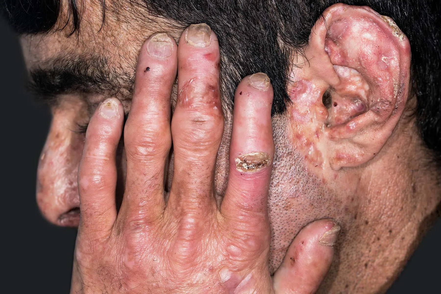
Cutaneous symptoms vary considerably among the different disorders. Porphyria cutanea tarda commonly affects the backs of the hands, forearms, face, and other sun-exposed areas. Possible signs include:
- Fragile skin that tears after minor bumps
- Fluid-filled blisters
- Slow-healing sores, crusting, or scarring
- Darkening or lightening of affected skin
- Increased facial hair growth
- Thickened or hardened patches of skin
Erythropoietic protoporphyria and X-linked protoporphyria often behave differently. Exposure to visible light may quickly cause tingling, itching, burning, swelling, and intense pain, particularly on the hands and face. The pain can be substantial even when little or no blistering is visible. Children may cry, withdraw their hands, or avoid outdoor activities before anyone recognizes light as the culprit.
Repeated skin injury in severe childhood porphyrias can lead to infections, scarring, changes in facial features, or damage to the fingers. Certain protoporphyrias can also affect the liver and gallbladder.
When symptoms require emergency attention
Seek emergency care for severe unexplained abdominal pain accompanied by weakness, confusion, seizures, persistent vomiting, fainting, difficulty breathing, or rapidly worsening neurologic symptoms. A person with known acute porphyria should bring medical-alert information and a current list of medications considered safe for that specific disorder.
What Triggers Porphyria Symptoms?
A genetic enzyme deficiency may create susceptibility, but an additional factor often pushes the heme-production pathway into overdrive and triggers an acute attack. Common triggers include:
- Certain prescription, over-the-counter, or herbal medicines
- Fasting, crash diets, very low calorie intake, or rapid weight loss
- Infections, surgery, and significant physical stress
- Hormonal changes, especially during the menstrual cycle
- Smoking
- Heavy alcohol consumption
- Severe emotional or psychological stress
Medication safety is particularly important. A drug that is harmless for most people may stimulate liver enzymes involved in heme production and provoke symptoms in someone with acute hepatic porphyria. Patients should use an expert-reviewed porphyria medication database and consult their clinician or pharmacist before starting or stopping medicines. Randomly abandoning necessary treatment is not a clever loophole; it can create an entirely new medical problem.
Triggers for porphyria cutanea tarda may include excess iron, hepatitis C, HIV, smoking, alcohol, estrogen therapy, and certain liver conditions. Identifying and treating these contributing factors can be an essential part of achieving remission.
How Porphyria Is Diagnosed
Porphyria cannot be diagnosed reliably from symptoms alone. Abdominal pain, anxiety, vomiting, neuropathy, and photosensitivity have many possible causes. Diagnosis begins with biochemical testing that matches the patient’s symptoms, followed by more specific tests to identify the exact form.
Testing during a suspected acute attack
For severe unexplained neurovisceral symptoms, the usual first-line test is a random urine sample for porphobilinogen, ALA, and creatinine. Creatinine allows the laboratory to adjust for how concentrated or diluted the urine is.
Urinary porphobilinogen is markedly elevated during attacks of acute intermittent porphyria, hereditary coproporphyria, and variegate porphyria. ALA dehydratase deficiency porphyria is an important exception because ALA rises without a comparable increase in porphobilinogen.
The sample should ideally be obtained while the person is symptomatic and before hemin treatment. It should be protected from prolonged exposure to bright light. A fresh random sample is generally more practical than waiting for a 24-hour collection, which may unnecessarily delay care.
A small elevation in total urinary porphyrins is not enough to prove acute porphyria. Mild increases can occur with liver disease, medication effects, and other medical conditions. Markedly elevated ALA or porphobilinogen is much more meaningful in a symptomatic patient.
Testing for cutaneous porphyria
When photosensitivity, blistering, or unexplained skin fragility is the main problem, testing may include plasma porphyrins, urine porphyrin fractionation, red blood cell porphyrins, and fecal porphyrins. The pattern of elevated compounds helps specialists distinguish porphyria cutanea tarda from variegate porphyria, erythropoietic protoporphyria, and other forms.
Plasma fluorescence scanning and specialized red blood cell testing can be useful in selected cases. Because porphyria assays are complex, abnormal results may need confirmation through a laboratory experienced in porphyria testing.
Genetic testing and family screening
After biochemical evidence identifies a porphyria pattern, genetic testing can confirm the responsible gene variant. Testing may also help identify relatives who carry the same variant and would benefit from trigger education.
Genetic testing should not be used as a stand-alone explanation for nonspecific symptoms. Many people with a pathogenic variant never develop clinical porphyria. The most reliable diagnosis combines the clinical history, biochemical findings, and genetic results rather than asking DNA to do all the detective work by itself. Genetic counseling can help families understand inheritance, testing options, and the meaning of positive or negative results.
Porphyria Treatment
There is no single porphyria treatment because each subtype produces a different chemical imbalance. Management may involve stopping an acute attack, preventing recurrence, reducing excess porphyrins, protecting the skin, treating liver-related triggers, and monitoring long-term complications.
Treatment for an acute porphyria attack
Moderate or severe acute attacks are generally treated in a hospital. The medical team may stop triggering medicines, treat infections, correct dehydration and electrolyte abnormalities, control nausea and pain, monitor breathing, and evaluate muscle strength.
Intravenous hemin is the standard specific treatment for a significant acute attack. Hemin suppresses the liver’s production of ALA and porphobilinogen, helping interrupt the biochemical process that drives neurologic symptoms. Improvement often begins within several days, although recovery from severe nerve injury may take much longer.
Carbohydrate or glucose therapy can suppress heme-pathway activity to a lesser degree. It may be used for mild attacks or as temporary support while hemin is being prepared, but glucose should not delay hemin when progressive neurologic symptoms are present.
Seizures must be treated carefully because some antiseizure drugs can worsen acute porphyria. Physicians should select medications from reliable porphyria safety resources while also correcting low sodium or other contributing abnormalities.
Preventing recurrent acute attacks
Prevention begins with regular meals, adequate carbohydrate intake, avoidance of extreme diets, prompt treatment of infections, smoking cessation, moderation or avoidance of alcohol, and careful medication review.
Givosiran is a monthly injectable therapy approved in the United States for adults with acute hepatic porphyria. It reduces production of an early liver enzyme in the heme pathway, lowering the neurotoxic precursors associated with attacks. Because it can cause significant adverse effects, treatment requires specialist supervision and laboratory monitoring.
Some patients with recurrent attacks may receive scheduled hemin therapy. Hormone-related attacks require an individualized plan involving a porphyria specialist and, when appropriate, a gynecologist. Liver transplantation is reserved for rare, severe cases in which repeated attacks continue despite other treatments.
Treatment for porphyria cutanea tarda
Porphyria cutanea tarda is often highly treatable. The two main approaches are:
- Therapeutic phlebotomy: Blood is removed at scheduled intervals to reduce excess iron, which gradually lowers porphyrin production.
- Low-dose hydroxychloroquine or chloroquine: These medicines help mobilize stored porphyrins so the body can eliminate them. Doses used for PCT are carefully controlled because excessive dosing can injure the liver.
Treatment also addresses contributing factors such as hepatitis C, HIV, alcohol, smoking, excess iron, estrogen exposure, or liver disease. Most patients can achieve biochemical and clinical remission, although follow-up testing is needed to detect recurrence.
Treatment for erythropoietic protoporphyria
People with erythropoietic protoporphyria or X-linked protoporphyria need protection from visible light as well as ultraviolet radiation. Helpful measures may include densely woven clothing, hats, gloves, window films, and specially selected sunscreens. Ordinary sunscreen may provide incomplete protection because visible light, rather than ultraviolet light alone, can trigger symptoms.
Afamelanotide is an implant approved for adults with erythropoietic protoporphyria who have a history of phototoxic reactions. It increases melanin and can extend pain-free light exposure. Patients must continue using protective measures and receive appropriate skin monitoring. Liver function and protoporphyrin-related complications may also require ongoing surveillance.
Treatment for severe childhood cutaneous porphyrias
Congenital erythropoietic porphyria may require intensive light protection, wound care, treatment of infections, transfusion support, and management of anemia or spleen enlargement. In carefully selected patients with severe disease, hematopoietic stem cell transplantation can correct the underlying blood-cell enzyme defect and may be curative. This treatment carries substantial risks and requires evaluation at a highly experienced center.
Living With Porphyria
A practical management plan is as important as the prescription list. People with porphyria may benefit from carrying a medical-alert card, wearing a medical identification bracelet, keeping a written emergency plan, and giving every clinician an updated medication-safety list.
Regular follow-up may include kidney testing, liver tests, blood pressure monitoring, iron studies, porphyrin measurements, or imaging. Some acute hepatic porphyrias and porphyria cutanea tarda are associated with an increased risk of liver cancer, so specialists may recommend periodic liver surveillance based on age, disease activity, and individual risk factors.
Nutrition should be balanced rather than extreme. A diagnosis of acute porphyria is not permission to survive entirely on cupcakes, but prolonged fasting and aggressive low-carbohydrate dieting can be dangerous for susceptible patients. Weight-loss plans should be gradual and supervised.
Mental health care also matters. Unpredictable attacks, chronic pain, diagnostic delays, and restrictions around sunlight can contribute to isolation, anxiety, or depression. Counseling and rare-disease support communities can help patients explain their limitations without feeling as though they must deliver a medical-school lecture every time someone proposes a beach picnic.
Patient Experience: The Long Road From Symptoms to Answers
The following composite example reflects experiences commonly described in clinical resources and patient communities. It does not represent one identifiable individual.
Imagine a 29-year-old woman named Rachel who develops severe abdominal pain over several days. The pain is constant and intense, but an ultrasound and CT scan do not show appendicitis, gallstones, kidney stones, or another obvious explanation. She becomes nauseated, cannot sleep, and notices that her heart seems to be racing. After receiving fluids and anti-nausea medicine, she improves enough to go home.
Two months later, it happens again. This time, Rachel also experiences constipation, weakness in her thighs, and unusual anxiety. Because the symptoms appear around a stressful week at work, they are initially attributed to panic. The anxiety is real, but it is part of the attack rather than the entire explanation. That distinction matters. Telling a patient that symptoms are “just stress” may sound efficient, but efficiency and accuracy are not the same sport.
During a third episode, a clinician notices the combination of severe unexplained abdominal pain, rapid heart rate, muscle weakness, low sodium, and a recently started medication. A random urine sample is collected for ALA, porphobilinogen, and creatinine while Rachel is still symptomatic. The porphobilinogen level is markedly elevated. Additional biochemical tests and genetic analysis confirm acute intermittent porphyria.
The diagnosis brings mixed emotions. Rachel is relieved that the symptoms have a name and a measurable biochemical cause. She is also unsettled by the fact that the condition is genetic, uncommon, and potentially serious. A porphyria specialist reviews her medicines, discusses likely triggers, and creates an emergency plan. She learns not to fast, to approach weight loss gradually, and to check medications before taking them.
When another attack begins, Rachel recognizes the pattern earlier. She brings her medical-alert information to the emergency department, where clinicians obtain laboratory samples, correct her low sodium, control vomiting, and administer hemin. The attack is treated before severe neuropathy develops. The experience is still unpleasant, but it is no longer a nameless medical ambush.
Someone with cutaneous porphyria may travel a different path. Consider a child with erythropoietic protoporphyria who cries after a short walk outside but has little visible rash. Adults may assume the child dislikes heat or outdoor play. Eventually, the family notices that burning pain and swelling consistently follow light exposure. Specialized blood testing identifies elevated protoporphyrin.
After diagnosis, the family adjusts daily routines. The child wears protective clothing, rides in a vehicle with appropriate window protection, and participates in outdoor events at safer times. Teachers receive a written explanation so light avoidance is not mistaken for stubbornness. Treatment does not eliminate every limitation, but it replaces confusion with planning.
These experiences highlight a central lesson: porphyria management is not limited to stopping symptoms. It involves being believed, obtaining the correct test at the correct time, identifying the exact subtype, and translating a rare diagnosis into ordinary daily decisions. Which medicine is safe? Can surgery proceed? How should a skipped meal be handled? What should an emergency physician know?
A strong care plan answers those questions before the next crisis. Patients should keep copies of confirmed laboratory and genetic results, document previous effective treatment, and identify a porphyria center or specialist who can advise local clinicians. Family members may also consider counseling and targeted genetic testing when a disease-causing variant has been confirmed.
Living with porphyria may require more preparation than most people need. Yet preparation creates freedom. A person who understands triggers, carries an emergency plan, protects the skin appropriately, and receives regular monitoring can make informed choices instead of organizing life around fear of the next unexplained symptom.
Conclusion
Porphyria is a group of uncommon disorders caused by disruptions in heme production. Acute forms can produce severe abdominal, neurologic, cardiovascular, and psychiatric symptoms, while cutaneous forms cause painful photosensitivity, blistering, skin fragility, or scarring.
Because the symptoms overlap with many common illnesses, accurate diagnosis depends on targeted biochemical testing. Urine ALA and porphobilinogen are central to evaluating a suspected acute attack, while plasma, blood-cell, urine, and fecal porphyrin tests help investigate cutaneous disease. Genetic testing is most useful after biochemical evidence has established the likely type.
Treatment may include intravenous hemin, carbohydrate support, givosiran, therapeutic phlebotomy, low-dose hydroxychloroquine, afamelanotide, light protection, treatment of contributing liver conditions, and careful avoidance of triggers. With specialist care and an individualized plan, many people can reduce symptoms, prevent complications, and live well with porphyria.