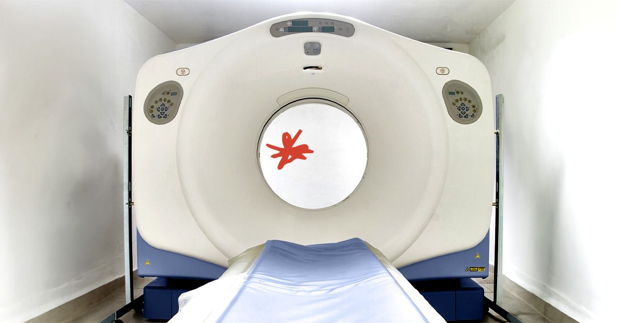
It is easy to imagine an MRI machine as a futuristic truth tunnel: you slide in, it hums like a spaceship with a grudge, and out comes a perfect map of everything happening inside your body. If only medicine were that tidy. The real answer to “Can an MRI detect cancer anywhere in the body?” is: MRI can detect many cancers in many parts of the body, but it cannot reliably detect every cancer everywhere, and it is not always the best first test.
Magnetic resonance imaging, better known as MRI, is one of the most powerful tools in modern cancer imaging. It can produce detailed pictures of soft tissues, organs, blood vessels, the brain, spine, pelvis, breasts, liver, pancreas, prostate, bones, and more. Doctors use it to find suspicious masses, check whether cancer has spread, guide biopsies, plan surgery or radiation, and monitor treatment response. But MRI does not “see cancer” the way a microscope sees cancer cells. It sees patterns: abnormal shapes, unusual tissue signals, swelling, contrast enhancement, restricted diffusion, and changes that may suggest a tumor.
That distinction matters. An MRI can raise suspicion, narrow possibilities, and sometimes strongly point toward cancer. However, in many cases, a biopsy or additional test is still needed to confirm the diagnosis. MRI is a brilliant detective, but it is not the judge, jury, and final pathology report.
How MRI Works in Cancer Detection
MRI uses a strong magnetic field, radio waves, and a computer to create detailed cross-sectional images of the body. Unlike X-rays and CT scans, MRI does not use ionizing radiation. That makes it especially useful when repeated imaging is needed or when doctors want excellent soft-tissue detail without radiation exposure.
Different MRI sequences highlight different tissue features. Some sequences show anatomy clearly. Others show fluid, fat, blood flow, inflammation, or how water molecules move inside tissue. Diffusion-weighted imaging, for example, can help identify areas where cells are packed tightly together, a pattern that can appear in some tumors. Contrast-enhanced MRI uses gadolinium-based contrast material injected into a vein to make blood vessels, inflammation, and some tumors stand out more clearly.
This is why MRI can be excellent for certain cancers. A small prostate tumor, a brain lesion, a liver mass, a soft-tissue sarcoma, or cancer spread in the spine may be much more visible on MRI than on a basic X-ray or even some CT scans. MRI is especially good at showing contrast between normal and abnormal soft tissue, which is its medical superpower. If CT is a detailed blueprint, MRI is the moody high-definition portrait that shows texture, depth, and suspicious shadows.
Can MRI Detect Cancer Anywhere?
MRI can image almost every region of the body, and whole-body MRI can scan large areas from head to thigh or head to toe. But imaging “anywhere” is not the same as detecting “everything.” Some cancers are too small, too flat, too early, too scattered, or too similar to normal tissue to be reliably found by MRI.
For example, MRI can be extremely useful for the brain, spinal cord, liver, pancreas, uterus, ovaries, prostate, bones, muscles, and soft tissues. It can also be used for breast cancer screening in people at high risk, usually alongside mammography rather than instead of it. In cancer staging, MRI may help determine whether a tumor has invaded nearby structures or whether disease has spread to bones or organs.
However, MRI is not ideal for every cancer type. Lung cancer screening is usually done with low-dose CT in eligible high-risk adults, because CT is better suited for finding small lung nodules. Colon cancer screening relies on colonoscopy, stool-based tests, and other targeted methods because early colon cancers or precancerous polyps may grow along the lining of the bowel and may not be obvious on a standard MRI. Skin cancers are usually detected by visual exam and biopsy. Leukemia and many blood cancers are diagnosed primarily through blood tests, bone marrow tests, and molecular studies, not by asking an MRI to patrol the bloodstream like a tiny medical submarine.
Where MRI Is Especially Helpful for Cancer
Brain and Spinal Cord Tumors
MRI is often the preferred imaging test for tumors in the brain and spinal cord. It provides detailed views of nervous system structures and can show swelling, tumor borders, pressure effects, and contrast enhancement. It is also commonly used to look for brain metastases in people with cancers that may spread to the brain, such as lung cancer, melanoma, breast cancer, and kidney cancer.
Liver, Pancreas, and Abdominal Tumors
MRI can be very useful for evaluating suspicious liver lesions, pancreatic abnormalities, bile duct tumors, kidney masses, adrenal lesions, and pelvic tumors. Specialized MRI techniques can help doctors tell whether a liver spot is likely benign, such as a hemangioma, or more concerning. In some situations, MRI gives information that CT or ultrasound cannot provide as clearly.
Prostate Cancer
Multiparametric prostate MRI has become an important tool in prostate cancer care. Doctors may use it to identify suspicious areas, guide targeted biopsy, evaluate cancer extent, and help decide between active surveillance and treatment. A prostate MRI does not replace every biopsy, but it can make the biopsy smarter, which is always appreciated by everyone involved.
Breast Cancer in High-Risk Patients
Breast MRI is highly sensitive and may detect some cancers that mammography misses, particularly in high-risk patients or people with dense breast tissue. But breast MRI is usually considered supplemental. Mammography remains the standard screening test for most women, while MRI is added when the risk profile justifies it, such as certain inherited gene mutations or a strong family history.
Bone, Muscle, and Soft-Tissue Tumors
MRI is one of the best imaging tools for soft-tissue sarcomas, bone marrow involvement, spinal tumors, and cancer that affects muscles, nerves, or connective tissue. It can show the relationship between a tumor and nearby nerves, vessels, joints, and muscles, which is crucial for surgical planning.
Where MRI May Miss Cancer or Be the Wrong Tool
No imaging test is perfect. MRI can miss tiny cancers, cancers that do not create a strong signal difference, tumors hidden by motion, or disease located in areas that are difficult to image. The lungs, for example, are full of air, and air is not MRI’s favorite dance partner. CT usually performs better for small lung nodules.
Some cancers begin on the surface lining of organs, such as early stomach, colon, bladder, or cervical cancers. These may require endoscopy, colonoscopy, Pap testing, HPV testing, cystoscopy, or tissue sampling. MRI may later help stage the disease, but it may not be the best way to find the earliest abnormal cells.
MRI can also produce false positives. A benign cyst, scar tissue, inflammation, infection, fibroid, enlarged lymph node, or harmless anatomical variation may look suspicious. This can lead to follow-up imaging, extra tests, anxiety, and sometimes biopsies that ultimately show no cancer. In other words, MRI can occasionally shout “plot twist!” when the story is actually boring.
What About Full-Body MRI Scans?
Full-body MRI has become more popular in wellness clinics and preventive health marketing. The promise sounds irresistible: scan the body, find silent problems early, sleep better at night. For some people with very high cancer risk, such as certain inherited cancer predisposition syndromes, whole-body MRI may be recommended as part of a medical surveillance plan. It is also used in selected cancer patients to look for metastatic disease or monitor conditions such as multiple myeloma.
For the average healthy person with no symptoms, no major risk factors, and no concerning family history, routine full-body MRI screening is much more controversial. Major radiology experts have warned that there is not enough evidence to recommend total-body MRI screening for everyone. The issue is not that MRI is useless; it is that screening healthy people can uncover many incidental findings that may never cause harm but still trigger expensive and stressful follow-up.
A full-body MRI might find something important. It might also find a tiny cyst, a benign nodule, a harmless bone island, a mildly enlarged lymph node, or a “please follow up in six months” mystery blob that takes up permanent residence in your thoughts. Preventive medicine should reduce risk, not create a subscription plan for anxiety.
MRI With Contrast: Why Doctors Use It
Some MRI exams are done without contrast. Others use gadolinium-based contrast agents to make certain tissues more visible. Contrast may help identify tumors, show whether a lesion has a blood supply, outline inflammation, or distinguish scar tissue from active disease. In cancer imaging, contrast can be especially helpful for brain tumors, breast MRI, liver lesions, pelvic tumors, and post-treatment follow-up.
Gadolinium contrast is generally considered safe for many patients, but it is not casual glitter for the bloodstream. Doctors consider kidney function, pregnancy status, allergy history, prior reactions, and the purpose of the scan before using it. People with severe kidney disease may need special precautions or alternative imaging. Patients should always tell the imaging center about kidney problems, implanted devices, metal fragments, pregnancy, breastfeeding, severe claustrophobia, or previous contrast reactions.
Can MRI Tell Whether a Tumor Is Cancerous?
Sometimes MRI findings are highly suggestive of cancer. A radiologist may describe a mass as suspicious based on its shape, borders, internal structure, enhancement pattern, diffusion restriction, invasion into nearby tissue, or associated enlarged lymph nodes. In certain organs, MRI scoring systems help standardize risk assessment, such as BI-RADS for breast imaging or PI-RADS for prostate MRI.
Still, MRI usually cannot prove that a tumor is malignant. Many benign conditions can mimic cancer, and some cancers look deceptively mild. The final answer often requires pathology, meaning cells or tissue are examined under a microscope. That is why a doctor may order a biopsy after an MRI. The scan shows where the problem is; the biopsy reveals what the problem is.
MRI vs. CT, PET, Ultrasound, and Other Cancer Tests
Choosing an imaging test is not a beauty contest where MRI automatically wins because it sounds expensive and comes with dramatic machine noises. Each test has strengths.
CT scans are fast and excellent for many chest, abdominal, bone, and emergency evaluations. They are commonly used for cancer staging, especially in the lungs and abdomen. PET scans show metabolic activity and can help detect active cancer throughout the body, often combined with CT or MRI. Ultrasound is accessible, radiation-free, and useful for thyroid, liver, pelvic, breast, and testicular evaluations. Mammography remains central to breast cancer screening because it can detect tiny calcifications that MRI may not show as well. Endoscopy and colonoscopy directly visualize internal linings and allow biopsy or polyp removal.
The best test depends on the suspected cancer, symptoms, risk factors, age, medical history, and what question the doctor is trying to answer. “Do I have a tumor?” “Has it spread?” “Is treatment working?” and “Is this scar tissue or active cancer?” are different questions, and they may require different tools.
When a Doctor May Recommend MRI for Cancer
A doctor may recommend MRI when symptoms, exam findings, lab results, or another imaging test raises concern. For example, MRI may be ordered after an ultrasound finds a liver lesion, after a PSA test raises concern for prostate cancer, after neurological symptoms suggest a brain or spine problem, or after a mammogram shows something that needs more evaluation in a high-risk patient.
MRI may also be used after cancer has already been diagnosed. In that setting, it can help determine tumor size, local spread, involvement of nearby organs, and whether surgery or radiation can be planned safely. After treatment, MRI can help monitor for recurrence or measure response to chemotherapy, radiation, immunotherapy, or targeted therapy.
What to Expect During an MRI
An MRI usually takes 20 to 90 minutes, depending on the body part and whether contrast is used. You may be asked to remove jewelry, watches, piercings, hearing aids, removable dental work, and anything metallic. The machine is loud, so earplugs or headphones are often provided. Some centers offer music, although the soundtrack may still be interrupted by the machine’s famous “industrial woodpecker inside a washing machine” rhythm.
You will need to stay still because movement can blur the images. If you feel claustrophobic, tell your doctor before the appointment. Options may include an open MRI, a wider-bore scanner, relaxation techniques, or medication. If contrast is needed, an IV will be placed. After the scan, a radiologist interprets the images and sends a report to your doctor, who explains the results in context.
Important Limitations: Why “Normal MRI” Does Not Always Mean “No Cancer”
A normal MRI is reassuring, but it does not guarantee that cancer is absent everywhere in the body. No scan can do that. Very early cancers, microscopic disease, abnormal cells in the blood or bone marrow, and cancers outside the scanned area may not appear. Even a full-body scan may use protocols that are broad rather than highly detailed for each organ.
This is why evidence-based screening still matters. Colonoscopy, Pap and HPV testing, mammography, low-dose CT for eligible lung cancer screening, skin checks, genetic counseling for high-risk families, and age-appropriate exams are not replaced by MRI. The smartest cancer detection strategy is targeted, risk-based, and guided by a cliniciannot a random medical treasure hunt.
Practical Examples
Example 1: Suspicious Headaches
A person develops new neurological symptoms, such as seizures, weakness, or vision changes. A brain MRI may be appropriate because it can show tumors, swelling, bleeding, inflammation, or other causes. If a suspicious mass appears, further imaging, blood work, surgery, or biopsy may follow.
Example 2: High PSA Level
A man has a rising PSA test. A prostate MRI may identify suspicious areas and help guide a targeted biopsy. The MRI may reduce unnecessary sampling in some cases, but if the scan or clinical risk remains concerning, biopsy may still be needed.
Example 3: Healthy Person Wants a Full-Body Scan
A healthy person with no symptoms wants reassurance. A full-body MRI might find nothing, or it might find incidental findings that require follow-up. The better first step is often a primary care visit, family history review, standard screening plan, and discussion of personal risk.
So, Should You Ask for an MRI?
If you have symptoms, abnormal lab results, a strong family history, or a known cancer diagnosis, asking your doctor whether MRI is appropriate is reasonable. But requesting an MRI “just to check everything” may not be the most effective route. The right scan at the right time can be life-changing. The wrong scan at the wrong time can be confusing, costly, and emotionally exhausting.
Good medical care is not about using the fanciest machine first. It is about matching the test to the question. MRI is a remarkable tool, but even remarkable tools need a reason to show up for work.
Experiences Related to MRI and Cancer Detection
People who undergo MRI for possible cancer often describe the experience as a mix of relief, worry, curiosity, and impatience. The scan itself is usually painless, but the emotional weight can feel much heavier than the machine’s table. Many patients say the hardest part is not lying still during the thumping sounds; it is waiting for the results afterward. The phrase “radiologist report pending” has a special talent for turning normal minutes into suspiciously long minutes.
One common experience is the “follow-up spiral.” A person may have an ultrasound, CT scan, blood test, or physical exam that shows something unclear. The doctor orders an MRI for a better look. The MRI then gives more detail: maybe the lesion has smooth borders and looks benign, or maybe it has features that require biopsy. In the best-case scenario, MRI turns a vague fear into a manageable answer. Even when it does not provide the final diagnosis, it often helps doctors decide the next step more confidently.
Another frequent experience involves incidental findings. Someone gets an MRI for back pain and learns they have a small kidney cyst. Or they get a scan for headaches and discover a harmless sinus issue. Sometimes these findings are useful. Sometimes they are the medical equivalent of opening a closet and finding three boxes you forgot existed. Most incidental findings are not cancer, but they can still create anxiety until a doctor explains what they mean.
Patients with known cancer often experience MRI differently. For them, the scan may be part of staging, treatment planning, or surveillance. A breast cancer patient may have MRI to evaluate the extent of disease before surgery. A prostate cancer patient may use MRI results to guide biopsy or treatment decisions. A person treated for a brain tumor may return for repeated MRIs to monitor for recurrence. In these cases, MRI becomes part of a longer relationship with the healthcare team. The machine may not become lovable, exactly, but it becomes familiar.
Claustrophobia is another real-world issue. Some people feel uncomfortable inside the scanner, especially during longer exams. Practical steps can help: asking about a wide-bore MRI, using headphones, practicing slow breathing, keeping eyes closed before entering the scanner, or discussing mild sedation with a doctor when appropriate. Many imaging centers are used to anxious patients, and there is no trophy for pretending to be calm while your nervous system is performing jazz drums.
People also learn that MRI results are not always simple “yes or no” answers. Reports may use phrases like “indeterminate,” “probably benign,” “suspicious,” “recommend follow-up,” or “correlate clinically.” This language can feel frustrating, but it reflects medical reality. Imaging is one piece of the puzzle. Doctors combine MRI results with symptoms, physical exams, lab tests, family history, prior imaging, and sometimes biopsy.
The most helpful experience usually comes when patients ask clear questions: What exactly did the MRI show? Is cancer likely, unlikely, or uncertain? Do I need another scan, a biopsy, or routine follow-up? What symptoms should I report? Are there standard screening tests I should still complete? These questions turn a confusing report into a plan, and a plan is much easier to live with than a mystery.
Conclusion
An MRI can detect many cancers in many parts of the body, but it cannot detect every cancer anywhere with perfect accuracy. It is strongest when doctors need detailed images of soft tissues, the brain, spine, pelvis, prostate, liver, breast, bones, and complex anatomy. It is less useful as a universal screening tool for healthy people and may miss certain early, microscopic, lining-based, lung, skin, or blood-related cancers.
The best way to think about MRI is this: it is an advanced imaging tool, not a magic cancer detector. It can reveal suspicious findings, guide biopsies, help stage cancer, plan treatment, and monitor response. But it works best when used for the right patient, the right body part, and the right clinical question. If you are worried about cancer, talk with a healthcare professional about your symptoms, personal risk, family history, and evidence-based screening options. The goal is not to scan everything possible. The goal is to find the right answer with the least unnecessary stress along the way.
Note: This article is for general educational purposes only and does not replace medical advice, diagnosis, or treatment from a qualified healthcare professional.