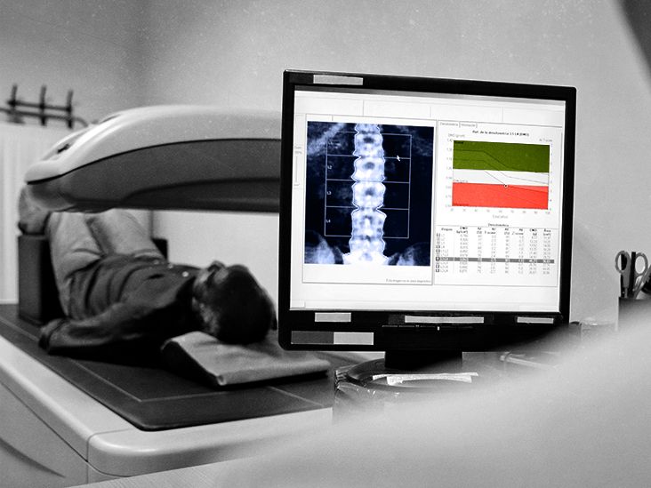
A bone density test can tell you a great deal about the strength of your bones, but it is not designed to diagnose arthritis. That distinction matters because osteoporosis and arthritis are frequently confused. Both can affect the skeleton, both become more common with age, and both occasionally appear in the same medical conversation. They are not, however, the same condition.
A dual-energy X-ray absorptiometry scanusually shortened to DXA or DEXAmeasures bone mineral density. Arthritis primarily affects joints, including cartilage, joint lining, nearby bone, tendons, and other supporting tissues. In other words, a DXA scan examines the strength of the building materials, while an arthritis evaluation looks at what is happening around the hinges.
The important twist is that arthritis can sometimes influence a bone density reading. A scan may contain clues suggesting degenerative changes, and certain forms of arthritis can increase the risk of osteoporosis. Understanding those connections can help you avoid treating one test result as an answer to an entirely different medical question.
The Direct Answer: A Bone Density Test Does Not Diagnose Arthritis
A standard bone density test does not confirm whether you have osteoarthritis, rheumatoid arthritis, psoriatic arthritis, gout, or another joint disease. Its primary purposes are to:
- Measure bone mineral density
- Identify osteopenia or osteoporosis
- Estimate the risk of future fractures
- Track changes in bone density over time
- Evaluate how well osteoporosis treatment is working
Arthritis is diagnosed through a different process. Depending on the suspected type, a health care professional may use your symptoms, medical history, physical examination, standard X-rays, blood tests, ultrasound, MRI, CT scans, or joint-fluid analysis.
So, if your knees ache when you climb stairs or your fingers feel stiff every morning, scheduling a DXA scan alone will not solve the mystery. It may provide useful information about your fracture risk, but your joints will still need their own investigation.
What Does a Bone Density Test Actually Measure?
A DXA scan uses two low-dose X-ray beams to estimate how much mineralprimarily calciumis present in a selected area of bone. Most central DXA examinations measure the lumbar spine and hip because fractures in these locations can have serious consequences.
The test is usually quick, painless, and noninvasive. You lie on a padded table while a scanning arm passes over part of your body. There are no tunnels, dramatic sound effects, or demands that you remain motionless for an entire geological era. Most people can return to normal activities immediately afterward.
Understanding the T-score
For postmenopausal women and men age 50 or older, results are commonly reported as a T-score. This compares your bone density with that of a healthy young adult reference population.
- -1.0 or higher: Generally considered normal bone density
- Between -1.0 and -2.5: Generally classified as low bone mass or osteopenia
- -2.5 or lower: Generally consistent with osteoporosis
A T-score is an important measurement, but it is not a complete fortune cookie for your skeleton. Clinicians may also consider your age, fracture history, medications, fall risk, family history, smoking, alcohol consumption, medical conditions, and calculated fracture risk.
Understanding the Z-score
A Z-score compares your bone density with that of people of similar age, sex, and body size. It is often emphasized in younger adults, premenopausal women, and children. An unexpectedly low Z-score may encourage a clinician to investigate medical conditions, nutritional problems, or medications that could be contributing to bone loss.
Can Arthritis Be Visible on a DXA Scan?
Sometimesbut visibility is not the same thing as diagnosis.
A DXA machine creates basic images so that the technologist can identify the bones being measured and analyze the correct regions. These images are not as detailed as ordinary diagnostic X-rays, MRI scans, or CT images. Nevertheless, significant osteoarthritis may produce visible structural changes, especially in the lumbar spine.
A radiologist or bone-density specialist might notice bone spurs, spinal degeneration, scoliosis, compression changes, surgical hardware, or other abnormalities. The report may mention such findings or recommend additional imaging. That observation does not mean the DXA scan has performed a complete arthritis evaluation.
Osteoarthritis Can Make Bone Density Look Higher
This is one of the most importantand least intuitiveconnections between arthritis and bone density testing.
Osteoarthritis can cause bone spurs, thickened bone, and other degenerative changes. In the lumbar spine, these extra-dense structures may be included in the DXA measurement. The machine can interpret them as additional bone mineral, making the spine’s bone density appear higher than it truly is.
Imagine weighing a suitcase after someone secretly added three hardback dictionaries. The scale is working correctly, but the number no longer represents the contents you intended to measure. Similarly, a DXA machine can accurately detect dense material while being unable to decide whether that density comes from healthy vertebral bone or degenerative changes around it.
When spinal arthritis significantly distorts the results, the interpreting clinician may exclude an affected vertebra, rely more heavily on hip or forearm measurements, compare the images with previous scans, or consider another method of evaluating bone strength.
What Tests Are Used to Diagnose Arthritis?
The appropriate tests depend on what type of arthritis is suspected. There is no universal arthritis test capable of identifying every condition with one heroic push of a button.
Tests for Osteoarthritis
Osteoarthritis is usually evaluated using symptoms, a physical examination, and standard X-rays. An X-ray may show narrowed joint spaces, changes in bone shape, increased bone density beneath damaged cartilage, cysts, or osteophytesbetter known as bone spurs.
Early cartilage damage may not appear clearly on an X-ray. MRI or ultrasound may be considered when symptoms are unexplained, soft-tissue damage is suspected, or more detail is needed.
Tests for Rheumatoid Arthritis
Rheumatoid arthritis is an autoimmune inflammatory disease. Diagnosis may involve:
- A review of joint pain, swelling, stiffness, and symptom patterns
- A physical examination of affected joints
- Blood tests for rheumatoid factor and anti-CCP antibodies
- Inflammation markers such as C-reactive protein and erythrocyte sedimentation rate
- X-rays, ultrasound, or MRI to evaluate inflammation and joint damage
No single blood test can diagnose every case of rheumatoid arthritis. Some people have the condition despite negative antibody tests, which is why the overall clinical picture matters.
Tests for Gout, CPPD, and Joint Infection
When gout, calcium pyrophosphate deposition disease, or an infected joint is suspected, joint-fluid analysis may be essential. A clinician removes a small sample of fluid with a needle and examines it for crystals, bacteria, and inflammatory cells.
A bone density scan cannot replace this process. Asking DXA to diagnose gout would be a little like asking a bathroom scale to identify what you ate for dinner. It measures something realjust not the thing you are trying to discover.
The Connection Between Rheumatoid Arthritis and Low Bone Density
Although a bone density test does not diagnose rheumatoid arthritis, people with rheumatoid arthritis may have a stronger reason to undergo bone-density testing.
Chronic inflammation can accelerate bone loss. Joint pain and fatigue may also reduce physical activity, depriving bones of the weight-bearing stimulation that helps maintain strength. In addition, corticosteroids such as prednisonesometimes used to control inflammatory diseasescan weaken bone when taken at sufficient doses or for extended periods.
Other factors may join the party without being invited: menopause, older age, low body weight, smoking, inadequate nutrition, vitamin D deficiency, and a previous fracture can all contribute to osteoporosis risk.
For someone with rheumatoid arthritis, therefore, two separate conditions may need attention:
- The inflammatory disease affecting the joints
- Loss of bone strength that increases fracture risk
Treating joint inflammation does not automatically correct osteoporosis, and osteoporosis medication does not control rheumatoid arthritis. Each condition needs an appropriate assessment and management plan.
Can You Have Arthritis and Osteoporosis at the Same Time?
Yes. Arthritis and osteoporosis can coexist, and their symptoms can occasionally be confused.
Osteoarthritis may cause pain, stiffness, reduced range of motion, and swelling around a joint. Osteoporosis, in contrast, usually causes no symptoms until a fracture occurs. A vertebral compression fracture can cause sudden back pain, gradual loss of height, or a stooped posture. Hip and wrist fractures may follow relatively minor falls.
A person could therefore have arthritic back pain, osteoporosis without symptoms, or both conditions simultaneously. Assuming that every ache comes from “weak bones” can delay an arthritis diagnosis. Assuming all pain is “just arthritis” can also allow an osteoporosis-related fracture to go unnoticed.
Clues That Deserve Prompt Medical Attention
Seek medical evaluation if you experience:
- Sudden severe back, hip, or groin pain
- Pain after a minor fall or ordinary movement
- New loss of height or a more rounded upper back
- A hot, red, intensely swollen joint
- Joint symptoms accompanied by fever
- Persistent morning stiffness or swelling in several joints
- Difficulty bearing weight on a leg
These symptoms do not prove arthritis or osteoporosis, but they warrant a closer look rather than a home diagnosis powered by coffee and search-engine optimism.
Who Should Consider a Bone Density Test?
In its 2025 recommendation, the U.S. Preventive Services Task Force recommended osteoporosis screening for women age 65 or older. It also recommended screening postmenopausal women younger than 65 when clinical risk assessment indicates increased fracture risk. For men, the Task Force concluded that evidence was insufficient to determine the overall balance of benefits and harms of population screening.
Individual medical organizations and clinicians may recommend testing in additional circumstances. A health care professional may consider a DXA scan when someone has:
- A fragility fracture
- Long-term corticosteroid exposure
- Rheumatoid arthritis or another condition associated with bone loss
- Early menopause
- Significant height loss
- Low body weight
- A strong family history of osteoporosis or hip fracture
- An abnormality seen on another imaging test
- A need to monitor osteoporosis treatment
Age alone should not be the only consideration. Your overall fracture risk and medical history matter more than whether your birthday cake has reached a particular number of candles.
How Arthritis Can Complicate DXA Interpretation
Accurate interpretation involves more than printing the lowest number on the page. The technologist and interpreting clinician must assess positioning, image quality, anatomical landmarks, previous surgery, implants, fractures, and structural abnormalities.
Conditions that may interfere with spinal measurements include:
- Osteoarthritis and large osteophytes
- Vertebral compression fractures
- Scoliosis or substantial spinal rotation
- Spinal fusion hardware
- Calcium deposits in nearby blood vessels
- Prior contrast material or other dense artifacts
This is why comparing a lumbar-spine T-score with a hip T-score can be revealing. If the spine appears unusually dense while the hip shows substantial bone loss, degenerative changes may be influencing the spinal result.
Do not attempt to “correct” the number yourself. A clinician may decide that certain vertebrae should be excluded or that another skeletal site provides a more reliable measurement. DXA interpretation is a medical analysis, not fantasy football for lumbar vertebrae.
Questions to Ask After Receiving Your Results
If your bone density report mentions arthritis, degenerative changes, or an unexpected difference between the hip and spine, consider asking:
- Was any part of my scan affected by osteoarthritis or another artifact?
- Which measurement is most reliable in my case?
- Do my results show normal bone density, osteopenia, or osteoporosis?
- What is my estimated fracture risk?
- Do I need a standard X-ray or another test for my joint symptoms?
- Could any of my medications be contributing to bone loss?
- Should I be evaluated for rheumatoid arthritis or another inflammatory condition?
- When should the bone density test be repeated?
Bring previous DXA reports whenever possible. Comparisons are most reliable when scans are performed using compatible equipment and analyzed with consistent methods. A small numerical change does not always represent genuine bone gain or loss.
Experiences Related to Bone Density Testing and Arthritis
The following representative experiences illustrate situations commonly encountered in medical practice. They are not descriptions of specific identifiable patients and should not be treated as substitutes for individual medical advice.
Experience 1: “My Spine Result Looked Great, but My Hip Did Not”
An older adult receives a DXA report showing normal-looking bone density in the lumbar spine but low density at the hip. At first, the spine result seems like excellent news. The patient jokes that at least one body part is overachieving.
On closer review, however, the DXA image shows significant lumbar osteoarthritis and several large bone spurs. Those dense degenerative changes have probably increased the measured spinal value. The clinician places more emphasis on the hip result and explains why the apparently strong spine score may be misleading.
The practical lesson is that the highest number is not automatically the most trustworthy one. Image quality and structural changes must be considered alongside the numerical score.
Experience 2: Joint Pain Leads to Two Separate Evaluations
A postmenopausal woman develops knee pain and assumes that a scheduled bone density test will reveal whether she has arthritis. Her DXA scan identifies low bone mass, but it does not explain the knee symptoms.
A separate weight-bearing knee X-ray shows osteoarthritis. She now has two related but distinct health concerns: reduced bone density and degeneration of the knee joint. Her treatment plan addresses fracture prevention, exercise, muscle strength, body mechanics, and management of knee pain.
This situation demonstrates why one imaging test cannot answer every musculoskeletal question. The DXA scan evaluates fracture risk; the knee X-ray evaluates the painful joint.
Experience 3: Rheumatoid Arthritis Raises a Bone-Health Question
A younger adult with rheumatoid arthritis has taken corticosteroids during several prolonged flares. The joints are the obvious concern, but the rheumatologist also asks about fractures, menstrual or hormonal history, calcium intake, vitamin D, activity level, and family history.
A DXA scan is orderednot to prove that rheumatoid arthritis exists, but to evaluate whether chronic inflammation and medication exposure have affected bone strength. The result shows low bone density, prompting a broader prevention plan.
For this patient, bone-density testing is part of managing the consequences and risks associated with inflammatory disease. It is not the diagnostic test for the arthritis itself.
Experience 4: A Normal DXA Does Not Cancel Persistent Joint Symptoms
A patient with morning hand stiffness receives a normal DXA result and assumes that nothing serious could be wrong. Months later, swelling develops in several finger and wrist joints. Blood tests, examination findings, and ultrasound ultimately support a diagnosis of inflammatory arthritis.
The normal bone density scan was not incorrect. It answered the question it was designed to answer: the patient’s measured bone mineral density was not low. It simply did not evaluate joint-lining inflammation.
This is an important reminder for anyone who receives a reassuring test result. “Normal” applies only to what was tested. Persistent swelling, prolonged morning stiffness, symmetrical joint symptoms, or declining function deserves appropriate follow-up even when bone density is normal.
What These Experiences Have in Common
Across these scenarios, the central theme is test selection. A DXA scan, standard X-ray, MRI, ultrasound, blood test, and joint-fluid analysis each offers a different type of information. The best test is not the most expensive or technologically impressive one; it is the test that answers the clinical question.
Patients often feel frustrated when one appointment produces another test instead of an immediate answer. Yet separating bone strength from joint disease prevents misleading conclusions. It also allows clinicians to recognize when both arthritis and osteoporosis require attention.
Conclusion
Does a bone density test show arthritis? Not in the diagnostic sense. A DXA scan measures bone mineral density and helps identify osteopenia, osteoporosis, and fracture risk. It does not provide a complete evaluation of cartilage loss, joint inflammation, autoimmune disease, gout, or other causes of arthritis.
However, arthritis may still affect the scan. Osteoarthritisparticularly in the lumbar spinecan create dense bone spurs and degenerative changes that make measured bone density appear artificially high. Rheumatoid arthritis can influence the picture in the opposite direction by increasing the risk of bone loss through inflammation, reduced activity, and corticosteroid use.
If you have joint pain, swelling, or stiffness, ask whether you need arthritis-specific testing. If you have risk factors for osteoporosis, ask whether a DXA scan is appropriate. The two evaluations may overlap, but they are not interchangeableand your skeleton deserves better than a one-test-fits-all strategy.