Wisdom teeth have a wonderfully misleading name. They do not improve your judgment, help you understand taxes, or stop you from texting an ex at midnight. They are simply the last permanent teeth to develop, usually appearing around the transition from the teenage years into adulthood.
Also called third molars, wisdom teeth sit at the farthest corners of the upper and lower jaws. Some grow in straight and behave like ordinary molars. Others arrive sideways, remain trapped beneath the gums, collect bacteria, or lean into neighboring teeth as though personal space were merely a suggestion.
Understanding what wisdom teeth look like, how they erupt, and which symptoms deserve attention can make dental decisions far less mysterious. Most importantly, having wisdom teeth does not automatically mean having surgery. Their position, health, function, and long-term risks all matter.
What Are Wisdom Teeth?
Wisdom teeth are the third and final set of molars. When all four develop, there is one at the back of each side of the upper jaw and one at the back of each side of the lower jaw. However, not everyone develops all four. A person may have one, two, three, four, or no wisdom teeth at all.
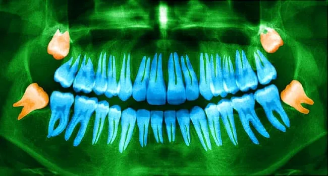
These teeth usually begin forming beneath the gums years before they erupt. Their crowns and roots continue developing during adolescence and early adulthood. A dental examination and X-ray can show whether they exist, how they are angled, whether their roots are fully formed, and how close they are to structures such as the lower jaw nerve or the maxillary sinus.
Why Do We Have Wisdom Teeth?
Wisdom teeth are generally viewed as an evolutionary leftover. Human ancestors had larger jaws and ate rougher foods that required more heavy-duty chewing. Extra molars were useful when meals involved tough plants, uncooked meat, and considerably fewer smoothies.
Modern food preparation has made eating easier, while human jaw size has changed over time. Many people now have limited room for a final row of molars. That mismatch between tooth development and available jaw space helps explain why third molars so often become impacted or erupt at awkward angles.
What Do Wisdom Teeth Look Like?
A fully erupted wisdom tooth usually resembles the other molars in the mouth. It has a broad chewing surface with grooves, ridges, and rounded cusps. The crown is typically white or ivory, while the roots extend into the jawbone beneath the gumline.
Compared with front teeth, molars look wide and somewhat blocky. Wisdom teeth can be less predictable than first and second molars, however. Their crowns may be smaller or irregularly shaped, and their roots may be straight, curved, fused together, or spread apart.
What a Healthy Erupted Wisdom Tooth Looks Like
A healthy wisdom tooth has emerged completely through the gum, sits upright, meets an opposing tooth appropriately, and can be brushed and cleaned around its entire surface. The surrounding gum should appear firm rather than swollen, bright red, or covered by a loose flap.
Because the tooth is located so far back, viewing it in a bathroom mirror may require impressive lighting and the jaw flexibility of a cartoon character. A dentist can examine it more accurately and check for hidden decay, gum pockets, or damage to the tooth beside it.
What a Partially Erupted Wisdom Tooth Looks Like
A partially erupted wisdom tooth may appear as a small white corner or bump breaking through the gum behind the second molar. Part of the chewing surface may be visible while the rest remains covered by tissue.
A flap of gum, called an operculum, can sometimes lie over the tooth. Food particles and bacteria may become trapped underneath it. The area can then become sore, swollen, difficult to clean, or infected. Inflammation around a partly erupted tooth is known as pericoronitis.
What an Impacted Wisdom Tooth Looks Like
An impacted wisdom tooth may not be visible in the mouth at all because it is partly or completely trapped beneath gum tissue or jawbone. Its position is usually revealed on a dental X-ray.
Common impaction patterns include:
- Vertical impaction: The tooth points upward but lacks enough room to erupt fully.
- Mesial impaction: The tooth tilts forward toward the second molar.
- Distal impaction: The tooth angles toward the back of the mouth.
- Horizontal impaction: The tooth lies almost sideways and may press directly against the neighboring molar.
Impacted wisdom teeth are common and may remain painless. Nevertheless, some can contribute to infection, cavities, gum disease, damage to nearby teeth, or cyst formation. Their appearance on an X-ray is only part of the decision-making process; symptoms, disease, age, anatomy, and surgical risk must also be considered.
When Do Wisdom Teeth Come In?
Wisdom teeth commonly erupt during the late teens or early twenties, although there is substantial variation. Some emerge later, some remain buried indefinitely, and others never form.
Eruption may happen gradually. A person might notice pressure behind the second molar, gum tenderness, or a small white edge appearing through the tissue. Mild temporary discomfort can occur as the gum opens, but persistent pain, swelling, discharge, or difficulty opening the mouth deserves professional evaluation.
Symptoms of Problematic Wisdom Teeth
A healthy wisdom tooth may produce no symptoms. When there is insufficient space, infection, decay, or inflammation, possible warning signs include:
- Pain or pressure at the back of the jaw
- Red, tender, or bleeding gums behind the second molar
- Swelling in the jaw, cheek, or nearby gum tissue
- Bad breath or an unpleasant taste
- Difficulty chewing or opening the mouth fully
- Food repeatedly becoming trapped behind a molar
- Headache or discomfort radiating toward the ear or face
- Visible decay in the wisdom tooth or neighboring molar
- Fever, pus, or enlarged lymph nodes when infection is present
Jaw pain is not automatically caused by wisdom teeth. Temporomandibular joint disorders, cavities, gum infections, sinus problems, clenching, and other conditions may produce similar discomfort. A dentist should identify the source rather than blaming the newest tooth in the neighborhood without evidence.
Do All Wisdom Teeth Need To Be Removed?
No. A wisdom tooth may be retained when it is healthy, functional, positioned correctly, surrounded by healthy gum tissue, and accessible for daily cleaning. Disease-free teeth that remain in place should be monitored through dental examinations and imaging when clinically appropriate.
Removal is more likely to be recommended when a wisdom tooth is associated with repeated infection, tooth decay, gum disease, cysts, damage to the second molar, nonfunctional positioning, or another planned dental or jaw procedure. Professional recommendations can differ for symptom-free impacted teeth because no single choice is right for every patient.
The American Association of Oral and Maxillofacial Surgeons supports surgical management for third molars associated with disease or a substantial risk of disease. When neither is present, active clinical and radiographic surveillance is an accepted approach. The American Academy of Pediatric Dentistry similarly recommends monitoring retained impacted teeth for changes or new pathology.
Questions To Ask Before Agreeing to Removal
- Is the tooth currently diseased, or is removal preventive?
- Could it damage the second molar if left in place?
- Can the tooth and surrounding gum be cleaned adequately?
- How close are the roots to a nerve or sinus?
- What are the risks of surgery compared with continued monitoring?
- Would a second opinion be reasonable?
How Wisdom Teeth Are Examined
A dentist begins with a medical and dental history, asks about symptoms, and examines the back of the mouth. The evaluation may include gum health, decay, eruption status, tooth function, jaw movement, and the condition of neighboring molars.
A panoramic X-ray can capture the teeth and jaws in one image. It helps show impacted wisdom teeth, root shape, bone coverage, and their relationship to nearby anatomy. In selected complex cases, three-dimensional imaging may provide additional detail, especially when a lower wisdom tooth appears close to the inferior alveolar nerve.
What Happens During Wisdom Teeth Removal?
Wisdom tooth extraction is usually an outpatient procedure performed by a dentist or oral and maxillofacial surgeon. The exact process depends on whether the tooth has erupted and how deeply it is positioned.
A straightforward erupted tooth may be loosened and removed from its socket. For an impacted tooth, the clinician may make an incision in the gum, remove a small amount of bone, divide the tooth into sections, and lift out the pieces. The area is then cleaned, and stitches may be placed.
Anesthesia and Sedation
Local anesthesia numbs the surgical area. Sedation may also be offered to reduce anxiety and awareness, while general anesthesia may be considered for certain complex procedures or individual medical needs. Patients receiving deeper sedation generally need a responsible adult to drive them home and follow the surgeon’s fasting and medication instructions precisely.
The procedure often takes less than an hour, although timing varies with the number, position, and complexity of the teeth. Removing one fully erupted tooth is quite different from removing four deeply impacted molars with curved roots.
Wisdom Teeth Removal Recovery
Bleeding, swelling, jaw stiffness, and tenderness are expected during the first few days. Swelling often becomes more noticeable before it begins to improve. Many people return to school or desk work within several days, but complete healing of the extraction sites takes longer.
Postoperative instructions commonly include biting gently on gauze, using cold packs as directed, resting, drinking water, and choosing soft foods. Yogurt, scrambled eggs, mashed potatoes, soup that is not piping hot, applesauce, and smoothies eaten with a spoon are popular choices. A straw is generally avoided early in recovery because suction may disturb the protective blood clot.
Patients should follow individualized instructions for brushing and rinsing. Vigorous swishing, smoking, vaping, alcohol, hard foods, and strenuous exercise may interfere with early healing. Good oral hygiene remains important, but the surgical sites need gentle treatment rather than an enthusiastic toothbrush excavation project.
Pain Control After Extraction
Nonopioid medications are frequently effective for acute dental pain. Depending on a patient’s health history, a dental professional may recommend ibuprofen, acetaminophen, or a carefully timed combination. These medicines are not appropriate for everyone, so dosing should come from the treating clinician rather than a social-media comment written by someone named “DentalBro92.”
Opioids are not routinely necessary for every wisdom tooth extraction and carry meaningful risks. The National Institute of Dental and Craniofacial Research notes that wisdom tooth removal can be a first exposure to prescription opioids for teenagers and young adults. Patients should discuss nonopioid options, medication interactions, and safe disposal of unused prescriptions.
Possible Complications
Most patients recover without serious problems, but every surgery has risks. Potential complications include infection, prolonged bleeding, injury to a neighboring tooth, sinus communication after an upper extraction, temporary or rarely persistent altered sensation from nerve irritation, and dry socket.
Dry Socket
Dry socket occurs when the blood clot protecting the extraction site is lost, breaks down too early, or does not form properly. The underlying bone becomes exposed, resulting in intense pain that may radiate toward the ear or temple. Bad breath or an unpleasant taste can accompany it.
Dry socket usually requires professional care. A dentist or surgeon may clean the area and place a soothing medicated dressing. It is not the same thing as ordinary postoperative soreness, especially when pain becomes dramatically worse after initially improving.
When To Call the Dentist or Surgeon
Contact the treating office promptly for uncontrolled bleeding, worsening swelling after several days, fever, pus, severe or escalating pain, persistent numbness, difficulty swallowing, trouble breathing, repeated vomiting, or an allergic reaction. Breathing difficulty, rapidly spreading facial swelling, or other signs of a serious infection require urgent medical attention.
How To Care for Wisdom Teeth That Stay
Keeping wisdom teeth means keeping them clean. Use a soft-bristled toothbrush with fluoride toothpaste and angle it carefully toward the back surfaces. Floss, interdental brushes, or another dentist-recommended device can help clean between the second and third molars.
Regular examinations matter because the far-back position can hide early cavities and gum pockets. A wisdom tooth that felt perfectly cooperative at age 20 may develop problems later. Retention is not a “forget it exists forever” plan; it is an active monitoring plan.
Realistic Wisdom Teeth Experiences: What the Process Often Feels Like
Wisdom tooth experiences vary widely, which is why comparing recoveries can be both reassuring and confusing. One person may have a fully erupted upper wisdom tooth removed in minutes and eat a soft dinner that evening. Another may have four deeply impacted teeth removed and spend several days looking as though a squirrel has stored emergency supplies in both cheeks.
A common first experience begins during a routine dental visit. The patient feels fine, but a panoramic X-ray shows lower wisdom teeth leaning toward the second molars. The dentist explains that the visible teeth are only part of the story: the crowns may be trapped beneath bone, while the developing roots sit near a sensory nerve. Rather than scheduling surgery instantly, the patient receives a referral for a more detailed consultation.
At the consultation, many patients are surprised by how much planning happens before the procedure. The surgeon reviews medications, allergies, previous anesthesia experiences, medical conditions, and the X-ray. The discussion may cover whether all four teeth should be removed together, which type of anesthesia is suitable, how long the patient should avoid food, and who will provide transportation afterward.
The day of surgery is often less dramatic than anticipated. With local anesthesia, pressure and movement may be noticeable, but sharp pain should not be. Patients receiving sedation may remember very little. Afterward, the mouth feels numb, gauze sits over the extraction sites, and speaking clearly becomes an optional feature. This is generally not the ideal moment to record a philosophical video for future generations.
The first evening typically revolves around rest, medication schedules, gauze changes, and soft food. A caregiver may need to repeat instructions because sedated patients are not always reliable historians. Mild oozing is common. Swelling and jaw stiffness may become more noticeable during the next day or two before gradually easing.
Food creates its own adventure. Mashed potatoes seem exciting for approximately one meal. By day three, many patients begin dreaming about crunchy tacos with the emotional intensity of a reunion scene. Returning to firmer foods should be gradual and based on comfort and the surgeon’s instructions. Small particles such as seeds, popcorn hulls, and sharp chips are usually poor early choices because they can irritate or lodge in healing sites.
Another common experience is worrying that every sensation signals a complication. A little soreness, swelling, bruising, or stiffness can be normal. Pain that suddenly intensifies, a foul taste accompanied by fever, persistent heavy bleeding, or progressive facial swelling is different. Written postoperative instructions are valuable because late-night internet searches tend to transform ordinary tenderness into seventeen rare diseases before breakfast.
Some people keep their wisdom teeth and have an entirely uneventful experience. Their third molars erupt upright, remain cavity-free, and can be cleaned without difficulty. Their main responsibility is attending regular checkups and resisting the belief that a lack of pain guarantees a lack of disease.
Others experience repeated irritation around a partially erupted tooth. Symptoms may improve after cleaning or treatment and then return months later. In those cases, removal may feel less like an unnecessary procedure and more like ending a recurring argument with the same tiny, enamel-covered troublemaker.
The most useful lesson from these varied experiences is that wisdom tooth care is personal. The number of teeth, angle of impaction, root development, medical history, oral hygiene, and proximity to nerves or sinuses all influence the recommendation. A friend’s easy extraction does not guarantee an identical recovery, and a relative’s difficult surgery does not mean yours will be difficult. Individual imaging and professional evaluation provide far better guidance than family folklore.
Conclusion
Wisdom teeth look much like ordinary molars when they erupt normally, but their hidden positions can range from upright to completely horizontal. Some remain healthy and useful for life. Others become difficult to clean, inflamed, infected, decayed, or damaging to nearby teeth.
The decision to remove or monitor wisdom teeth should be based on symptoms, examination findings, imaging, function, disease risk, and the risks of treatment. A dentist or oral surgeon can explain what your teeth look like beneath the gums and help you choose a plan that makes sense for your mouthnot somebody else’s.